
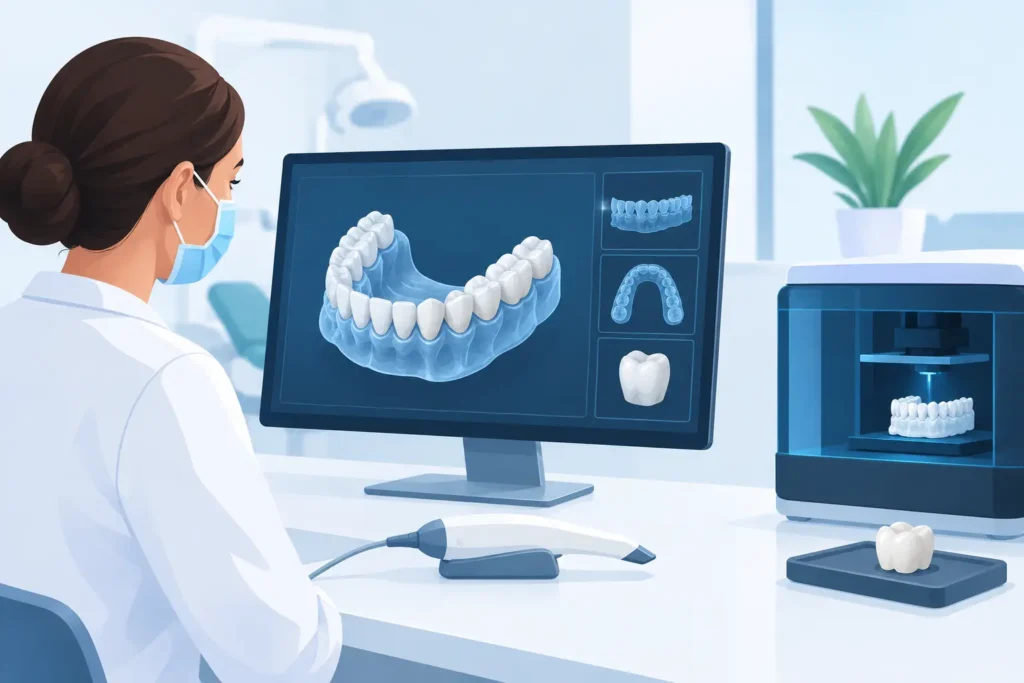
A full-arch case can lose momentum long before the final prosthesis is delivered. A marginal scan artifact, incomplete implant record, unclear smile reference, or late-stage change in vertical dimension can create avoidable adjustments and difficult patient conversations. Digital dentistry gives the clinical team and laboratory a clearer way to manage those variables from the first record to final delivery.
For dentists who routinely manage cosmetic, implant, and rehabilitative cases, the value is not simply faster production. The real advantage is better visibility into the treatment plan, more precise communication, and a workflow that supports fit, function, and lifelike esthetics with greater consistency.
What Digital Dentistry Changes in the Restorative Workflow
Digital dentistry connects clinical records, virtual design, manufacturing technology, and laboratory craftsmanship. An intraoral scan can replace or supplement conventional impressions, but scanning alone is not the workflow. The benefit comes from how each record is evaluated, designed, verified, and translated into a final restoration.
In a well-managed digital case, the scan, photographs, shade information, bite records, facial references, implant library data, and treatment goals are considered together. That gives the laboratory team a more complete picture of what the restoration must accomplish. For a single posterior crown, the priority may be contact integrity and occlusal function. For a veneer case or full-mouth rehabilitation, facial support, incisal edge position, phonetics, gingival architecture, and the patient’s expectations carry equal weight.
Digital tools make it easier to preserve and share this information. They do not eliminate clinical judgment. A scanner cannot determine whether a proposed incisal length supports the patient’s face, whether tissue is stable enough for the final record, or whether the functional plan is appropriate. Those decisions remain clinical decisions, strengthened by better records and closer laboratory collaboration.
Better Records Create Better Starting Points
The first major gain is diagnostic clarity. High-quality digital records give the dentist and technician an opportunity to identify concerns before fabrication begins rather than after the restoration arrives in the operatory.
For fixed restorative cases, scan quality directly affects margins, contacts, emergence profile, and occlusion. Tissue must be managed appropriately, preparations must be visible, and the captured bite must accurately represent the planned restorative position. A fast scan is not necessarily a usable scan. The goal is a clean, complete digital impression that provides enough information for the laboratory to work confidently.
For implant cases, the record becomes more demanding. Scan body seating, orientation, implant system selection, soft-tissue contours, opposing dentition, and verified jaw relationship all influence the final result. A minor discrepancy at the record stage can become significant across a multi-unit implant bridge. This is why complex digital cases benefit from deliberate verification protocols, not assumptions based on a screen image alone.
Digital Records Need Clinical Context
A digital file does not communicate everything a skilled technician needs to know. Photographs remain essential for esthetic work. Full-face, retracted, lateral, and shade images help guide line angles, translucency, texture, characterization, and tooth proportions. Diagnostic information such as planned changes to vertical dimension, provisional feedback, occlusal concerns, and patient priorities also needs to accompany the case.
The most productive cases begin with a conversation, not just an upload. When the laboratory understands the clinical objective, it can identify missing records early, recommend a design sequence, and help the team avoid costly remakes or chairside compromises.
Digital Design Improves Communication Before Production
The design phase is where digital dentistry becomes especially valuable for high-esthetic and complex restorative treatment. Virtual wax-ups allow the dentist to evaluate proposed tooth position and contour before irreversible preparation or final fabrication. This is particularly useful when changing length, closing spaces, rebuilding worn dentition, or planning an implant-supported rehabilitation.
A diagnostic design can support patient communication as well. Patients often understand treatment more readily when they can see a proposed change in tooth position or smile form. That clarity can improve acceptance, but it must be presented responsibly. A digital preview is a planning tool, not a guarantee that ignores biology, occlusion, periodontal limitations, or material requirements.
For the clinical team, the design provides a common reference point. The dentist can approve contours, functional space, and esthetic direction. The laboratory can build provisionals, matrices, reduction guides, or final restorations around the approved plan. When changes are needed, they can be discussed while they are still efficient to make.
The Provisional Still Does Essential Work
Digital planning does not diminish the value of well-made provisionals. In many comprehensive cases, the provisional is the patient’s functional trial period. It tests speech, comfort, occlusion, esthetic preferences, and the patient’s ability to maintain the planned result.
Feedback from the provisional phase should move back into the digital workflow. If the patient prefers a shorter incisal edge, if phonetics reveal a concern, or if functional adjustments change the occlusal scheme, those findings should inform the final design. The most predictable digital workflow is iterative when the case calls for it.
Precision Manufacturing Is Only Part of the Equation
Milled zirconia, lithium disilicate, printed models, digitally designed splints, and surgical guides can offer impressive accuracy and efficiency. Yet the material and manufacturing method should follow the clinical indication, not the other way around.
Zirconia may be an excellent choice for strength-driven posterior or full-arch applications, while layered ceramics may be selected when maximum anterior characterization is required. Lithium disilicate can offer a compelling balance of esthetics and conservative preparation in the right situation. Implant restorations may require careful material selection based on restorative space, load distribution, opposing dentition, hygiene access, and the design of the supporting framework.
The digital workflow improves repeatability, but premium outcomes still depend on experienced technicians who understand morphology, occlusion, ceramic behavior, implant prosthetics, and natural esthetics. A digitally milled restoration is not automatically a refined restoration. Design review, material handling, characterization, and quality control remain decisive.
At Mizrachi Dental Lab, digital production is supported by the same craftsmanship that has guided four decades of restorative work. The objective is not to replace technician expertise with technology. It is to apply advanced technology in a way that gives dentists more dependable fit, efficient case communication, and restorations that look natural in the patient’s smile.
Where Digital Dentistry Requires Extra Care
Digital workflows can reduce friction, but they are not the right shortcut for every clinical situation. Deep subgingival margins, uncontrolled bleeding, mobile tissue, limited opening, reflective surfaces, and challenging full-arch scans can make conventional records, verification records, or a combined analog-digital approach more appropriate.
Edentulous and complex implant cases deserve particular caution. Capturing soft tissue and an accurate maxillomandibular relationship in a fully edentulous patient can be more complex than scanning a dentate quadrant. Depending on the case, conventional records, photogrammetry, verification jigs, printed try-ins, or a staged record protocol may be necessary. The right workflow depends on the patient, the restoration design, the clinician’s experience, and the accuracy required at each stage.
The same principle applies to surgical guides. Digital planning can support more prosthetically driven implant placement, but guide accuracy depends on sound diagnostics, stable guide support, accurate imaging, correct implant library data, and disciplined surgical execution. Technology supports clinical judgment. It does not replace it.
Building a More Predictable Lab Partnership
The best digital results come from defined responsibilities. The practice should establish consistent scanning protocols, capture required photographs, document implant components accurately, and communicate the intended restorative outcome. The laboratory should review incoming records carefully, raise concerns early, provide design support when needed, and maintain quality standards through every production stage.
This level of coordination matters because fewer surprises mean more than fewer adjustments. It can protect chair time, improve patient confidence, reduce remake risk, and help the practice deliver the quality of care its reputation depends on. For larger cases, it also gives the entire clinical team a more organized path through planning, provisionalization, verification, and final delivery.
Digital dentistry is most valuable when it makes excellent dentistry easier to repeat. Start each case with complete records, a clear treatment objective, and a laboratory partner prepared to question details before they become complications. That discipline creates the conditions for better-fitting restorations, stronger patient trust, and results worth referring to others.